
Membrane proteins do not function in isolation. They operate within lipid bilayers, surrounded by water, ions, and other molecular components that influence their structure and biological activity.
For researchers in computational drug discovery, understanding this environment is essential. The position of a protein within the membrane, its interaction with surrounding lipids, and the accessibility of specific structural regions can provide valuable context for subsequent molecular studies.
However, visualizing a complete membrane system presents a practical challenge. A single molecular model may contain hundreds of thousands of atoms, making it difficult to distinguish the protein from its surroundings.
How can researchers explore these complex systems without losing important structural information?
Pauling.AI’s Structure Explorer allows researchers to inspect molecular assemblies, control the visibility of individual components, and adjust molecular representations within a single interactive environment.
In this example, we explore a single membrane protein system based on beta2 adrenoceptor (PDBID: 4LDE) to demonstrate how structural visualization can make complex molecular environments easier to interpret.
Understanding the membrane protein environment
Membrane proteins are involved in essential biological processes, including molecular transport, cellular signaling, and communication across membranes. Many are also important therapeutic targets.
Unlike soluble proteins, membrane proteins interact with lipid bilayers. Their transmembrane regions occupy the membrane interior, while other domains may extend into the surrounding aqueous environment.
A representative membrane model may contain the protein, two lipid leaflets, water molecules, ions, ligands, and cofactors.
These components provide structural context, but displaying them simultaneously can make the system difficult to navigate.
The challenge is therefore not simply visualizing more atoms. It is identifying which components are relevant to the question being investigated.
Visualizing a complete membrane system
The Structure Explorer provides an interactive three-dimensional environment for examining molecular systems.
The loaded 4LDE-based membrane complex (membrane building by Pauling.AI) contains hundreds of thousands of atoms, including the β2-adrenergic receptor embedded in a lipid bilayer and surrounded by water and other molecular components (fig. 1).
The protein is displayed in turquoise, while the lipid bilayer appears in contrasting orange and yellow tones. Water molecules are represented as numerous points surrounding the membrane.
This representation helps establish the overall organization of the molecular assembly and the position of the protein relative to its environment.
The interface also provides options for changing molecular representations, adjusting opacity, and exploring the structure from different perspectives.
These controls allow researchers to inspect the same structural model at different levels of visual detail.
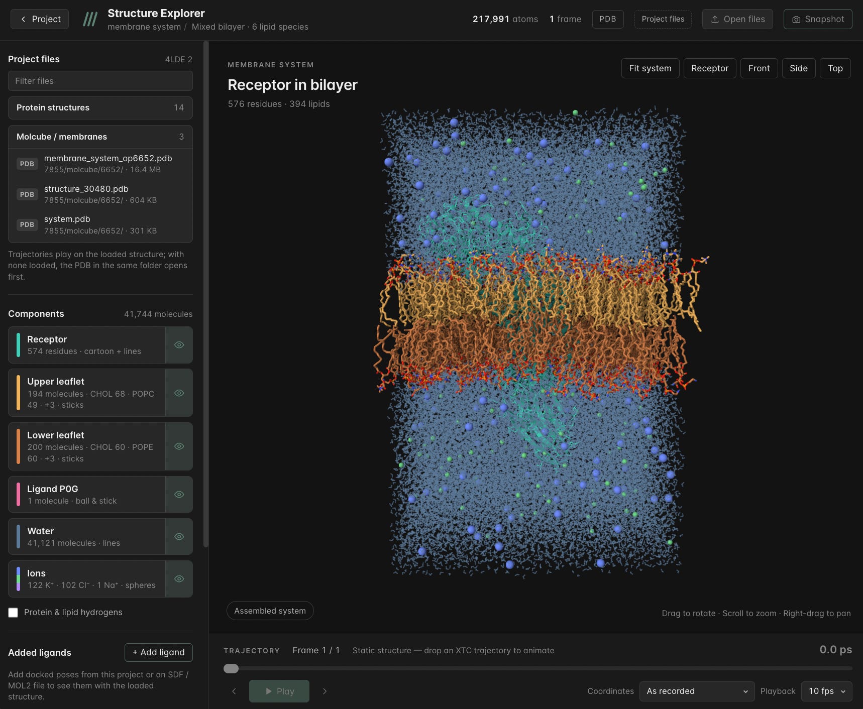
Figure 1. Structure Explorer GUI displaying the 4LDE-based membrane system and its molecular components in an interactive 3D view.
Isolating molecular components with one click
Displaying every molecular component is useful for understanding the complete system, but it can obscure specific structural regions.
One of the Structure Explorer’s practical features is the ability to control component visibility directly from the interface.
In another view of the same 4LDE-based molecular system, the receptor, upper and lower lipid leaflets, ligands and cofactors, and water are organized into separate component groups.
Each group can be displayed or hidden independently (fig. 2).
For instance, hiding water molecules reduces visual density and makes the relationship between the membrane protein and lipid bilayer easier to examine.
Importantly, this operation changes the visualization rather than the molecular structure itself. Researchers can simplify the displayed system without deleting atoms from the loaded model.
The component panel also provides a convenient way to switch between different structural views without manually editing molecular files.
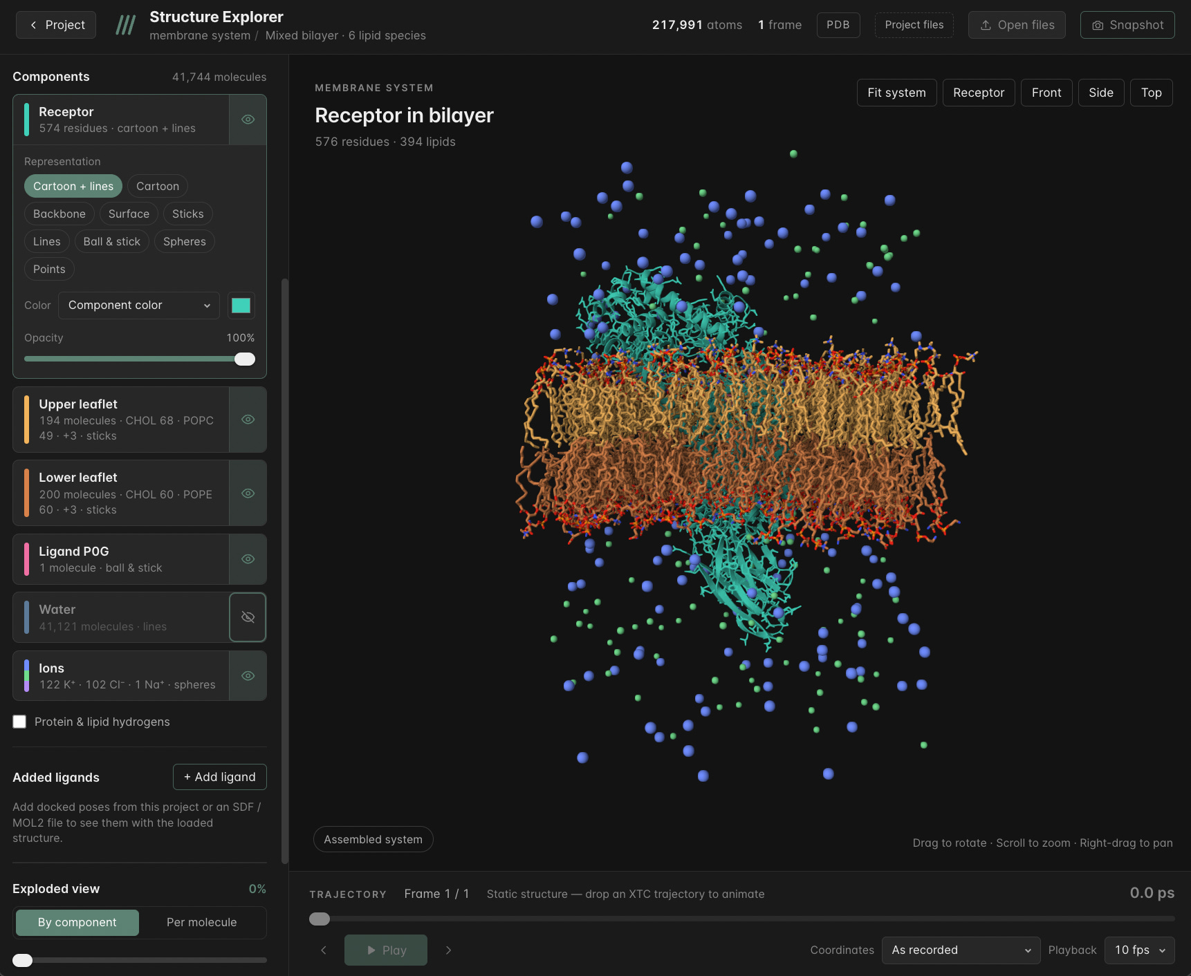
Figure 2. Component visibility controls in the Structure Explorer GUI, allowing users to show or hide molecular groups within the same 4LDE-based system.
Exploring the protein membrane interface
Once the surrounding water is hidden, the relationship between the protein and membrane becomes easier to inspect.
Researchers can focus on how the protein is positioned relative to the lipid leaflets and distinguish regions located within the membrane from those extending into the surrounding aqueous environment.
The Structure Explorer also provides different viewing perspectives and molecular representation options, allowing users to inspect the same system in different ways.
For instance, a cartoon representation can make the protein’s overall architecture easier to follow, while alternative representations of the surrounding molecules can help reveal how the system is organized.
Rotating and zooming into the structure provides additional perspectives without requiring a separate molecular model.
These controls are useful when examining complex membrane assemblies, where the complete system and inividual components may each require a different level of visual detail (fig. 3).
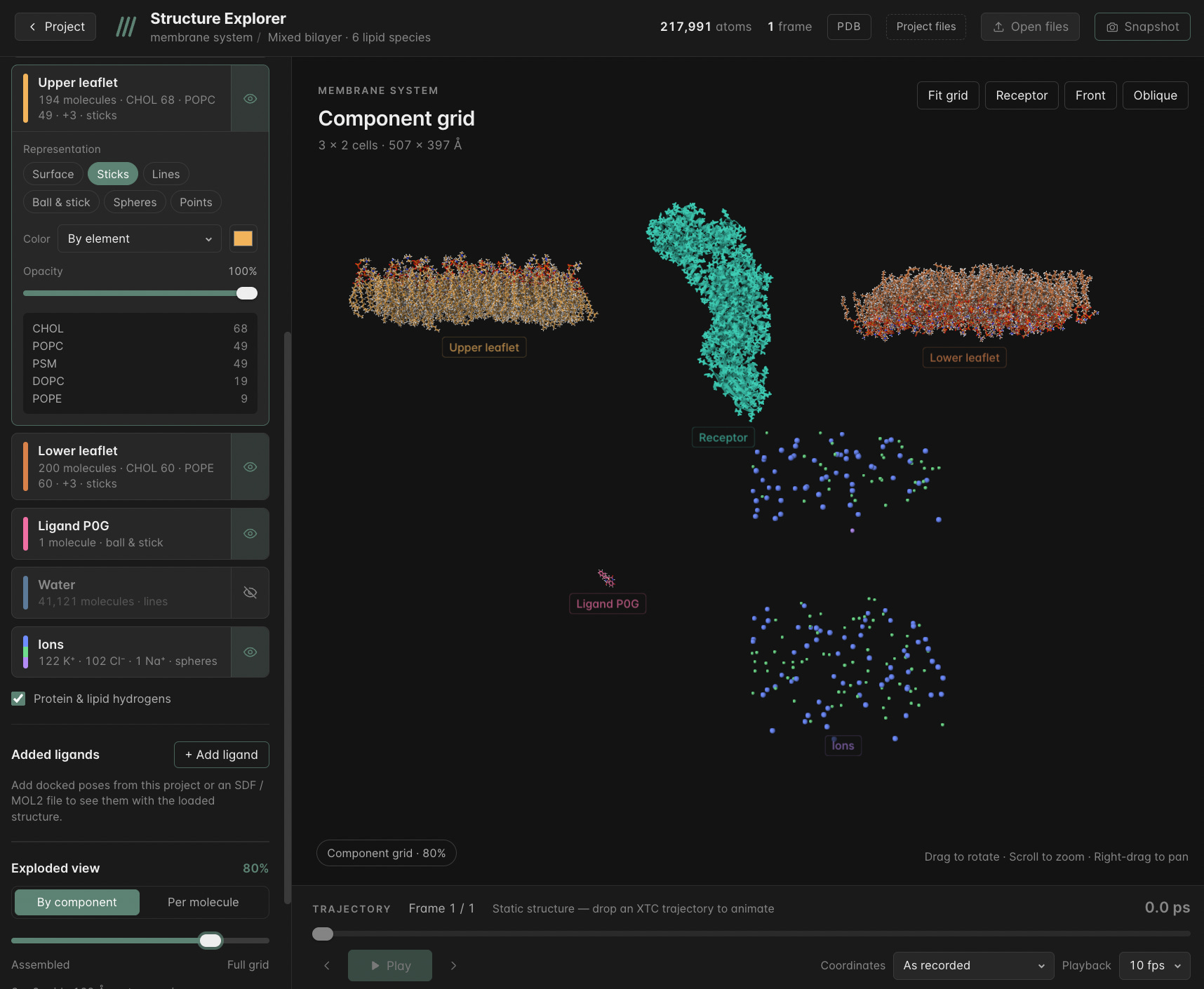
Figure 3. Structure Explorer GUI options for adjusting molecular representations and examining the 4LDE-based system from different perspectives.
Why structural visualization matters in drug discovery
Structural visualization supports several stages of computational drug discovery.
Before performing detailed molecular calculations, researchers often need to inspect protein orientation, identify relevant structural regions, and understand how the surrounding molecular environment is organized.
For membrane associated targets, this context can be particularly important because lipid interactions and membrane positioning may influence subsequent modeling decisions.
Interactive visualization can support these activities by making complex structures easier to inspect and communicate.
However, visualization alone does not establish binding affinity, molecular stability, or dynamic behavior. These questions require appropriate computational methods or experimental validation.
Instead, the Structure Explorer provides a practical starting point for structural interpretation and the planning of more detailed investigations.
From molecular complexity to structural clarity
Molecular models containing hundreds of thousands of atoms provide valuable information, but interpreting them requires control over how that information is displayed.
The 4LDE-based membrane system presented here illustrates how researchers can move between a complete molecular assembly and focused structural views using Pauling.AI’s Structure Explorer.
By combining customizable representations, interactive navigation, and component-level visibility, the platform makes it easier to examine complex molecular environments without altering the underlying structural model.
Clearer visualization does not replace computational analysis, but it helps researchers understand the systems they are studying and determine where to investigate next.
Explore complex molecular systems with Pauling.AI.
References
Locher, K. P. (2009). Structure and mechanism of ATP-binding cassette transporters. Philosophical Transactions of the Royal Society B. https://doi.org/10.1098/rstb.2008.0125
A brief history of visualizing membrane systems in molecular dynamics simulations. (2023). Frontiers in Bioinformatics. https://doi.org/10.3389/fbinf.2023.1149744